How Can I Increase My Transfection Efficiency?

If you work in gene therapy, cell biology, or cancer research, cell transfection is likely a part of your daily routine. To that end, you want to ensure you have the highest rates of transfection efficiency.
Or, if you’re new to transfection, you might have stumbled here for some building blocks of proper techniques.
Whichever category you fall under, welcome! This guide will outline:
- A Brief Guide to Transfection
- What Determines Transfection Efficiency
- How to Increase Efficiency
So, if you’re already well-versed in transfection, transfection techniques, and what affects the efficiency—amazing!—go ahead and skip to the third section where you will learn how to improve your efficiency. Otherwise, let’s begin.
Part 1 – Hitchhiker’s Guide to Transfection
Welcome to the wonderful universe of nucleic acid-delivery systems (and the powerful analytical tools they provide as a byproduct). While one article clearly isn’t enough to explore every galaxy, we can point your telescope in the right direction. Below, let’s focus on the basics:
- What is transfection?
- What are the two introduction methods?
- Biological, chemical, and physical processes of transfection; what’s the difference?
Class is now in session.
What is Transfection?
Transfection is the process by which foreign nucleic acids (RNA, DNA, etc.) are transferred into a healthy cell, to produce a “genetically modified cell.” The term itself is a combination of “trans” and “infection,” which might glean some intuition into its operation.
Though it’s considered a technique, its far-reaching consequences mark it as more of a powerful, analytical tool. Cell transfection allows researchers to interact with, exchange, and hyper-analyze small chains of nucleic acids. Why is this so powerful?
Let’s use human DNA and basic arithmetic to consider this:
- Human genes are frequently found to be around 27,000 base pairs long, with some stretching closer to 2 million base pairs.
- The bible itself (King James Authorized Bible) has around 3 million letters (or 1.5 million letter pairs—though, that’s not how normal people think of reading).
- Meaning, trying to analyze just one protein-builder inside one gene involves reading a bible-length book made up of four letters (A, T, G, C) and analyzing individual pieces.
Tough problem to crack—enter, the transfection of cells.
During a transfection, foreign nucleic acid chains into a healthy cell. The nucleic acids then combine with the existing DNA in the cell, creating a genetically modified cell. Researchers can then study the changed properties of the cell. Because the cell nucleic acid sequence was known beforehand, researchers can further analyze the gene function and protein expression of the transfected cell.
What are the Two Introduction Methods?
The method mentioned above, while simplified, is only one type of nucleic acid introduction method: stable transfection. The other type, transient transfection, is an impermanent way to integrate genes.
- Stable Transfection – Foreign nucleic acids bypass the cell membrane, enter the cell, and are delivered through the nuclear membrane. From there, foreign nucleic acids integrate with the host DNA (or RNA) to create a modified gene.
- All future cell progeny will have this modified gene integrated
- Stable integration has a lower uptake (more rare occurrence)
- Includes a biomarker to calculate transfection efficiency
- Useful for drug screening, studying morphological changes, antibiotic resistance, and more
- Transient transfection – Foreign nucleic acids bypass the cell membrane, enter the cell, and (if DNA) are delivered through the nuclear membrane. Foreign DNA does not integrate into the host DNA, but continues to express itself through protein production.
- Foreign DNA will not pass on to future cell progeny
- Environmental factors and cell division will end the expression
- Useful for understanding gene silencing, inhibitory RNA, and small-scale protein production
Which transfection method you choose for your experiment heavily depends on two factors: what you’re measuring and what cell population you’re using.
Related: How to Complete Cell Cycle Analysis via Flow Cytometry
Processes of Transfection | Biological, Chemical, Physical
As a final part of this introductory course, let’s break down some of the different processes by which transfection of cells can occur. This will provide the necessary baseline understanding of the different factors of cell transfection efficiency (in Part 2).
- Biological Transfection – The common method for clinical research involves transfecting via viruses. Often referred to as transduction, virus-mediated transfection infects the cell much like a normal virus and integrates the DNA into the host nucleus for replication.
- Chemical Transfection – Contemporary research often uses polymers and lipids to carry nucleic acid chains into the cell. By negatively charging the cell, and positively charging the polymers, these two are attracted and eventually, the nucleic acid is deposited in the cell.
- Physical Transfection – Laser based transfection is often what’s thought of for physical methods. In some cases, such as phototransfection, laser beams generate holes at a specific subcellular location to allow for the foreign nucleic acids to be delivered.
With the basics now thoroughly ingrained into your noggin, let’s dive into the juicy details about transfection efficiency.
Part 2 – What Determines Transfection Efficiency
While each transfection procedure listed above will come with its own advantages and disadvantages, as well as its own set of techniques to improve the efficiency rate, there are overall factors that contribute. This high-level analysis is what will be most helpful here. To that end, let’s discuss cell transfection efficiency:
- The health and viability of the cell population (very important!) – In the process of creating cell assays, cells are sent through the ringer. And if you don’t have a healthy cell population, you’re guaranteed to not just have a low transfection efficiency, but also incorrect and misleading results. You can see why cell viability is such a big factor.
- Cell confluency – Confluency measures how packed the cells are on a given surface (not to be confused with cell density). A 50% cell confluency means that half the surface is covered. Too little or too much confluency will affect how receptive the cell is to transfection.
- Type of cell – Cells have different life spans, cell wall characteristics, and functions. As such, some cells will be easier to perform transfection, changing the efficiency.
- Type of nucleic acid transfected – Similarly to the different types of cells, nucleic acids range in complexity. DNA is more complex than RNA which is more complex than some individual nucleic acid chains. In general, the more complex, the more difficult the transfection process is.
- Method of transfection – Depending on the biological, chemical, and physical transfection method, you’ll have a range of efficiencies to strive for.
Part 3 – How to Increase Transfection Efficiency
Within the different processes (biological, chemical, physical) there are continual improvements in technology and technique. For example, every advancement in optic technology and laser precision is going to have a beneficial impact on photo transfection.
Similarly, once we create the shrink ray, we can hand deliver these nucleic acids and use our equally tiny screwdrivers to lock the nucleic acids in the correct place, all without harming the cell.
But until then, here the accepted methods of increasing the cell transfection efficiency are:
- Transfecting healthy cells that are still actively dividing
- Optimizing the nucleic acids (both high-quality and proper amount)
- Correcting for cell population per individual well
Healthy Cells Are Happy (and high transfection-rate) Cells
As mentioned above, cells that are separated and counted are given the short end of the cellular stick. Cell viability is incredibly important. Within industry-standard flow cytometers, they are exposed to excessive pressures that cause wear and tear to the cell wall, as well as damage the internal organelles. They are pushed and prodded and, in the end, experimented on. Without anthropomorphizing these individual cells (aka, don’t feel bad for them), it’s easy to see why this won’t produce the best results.
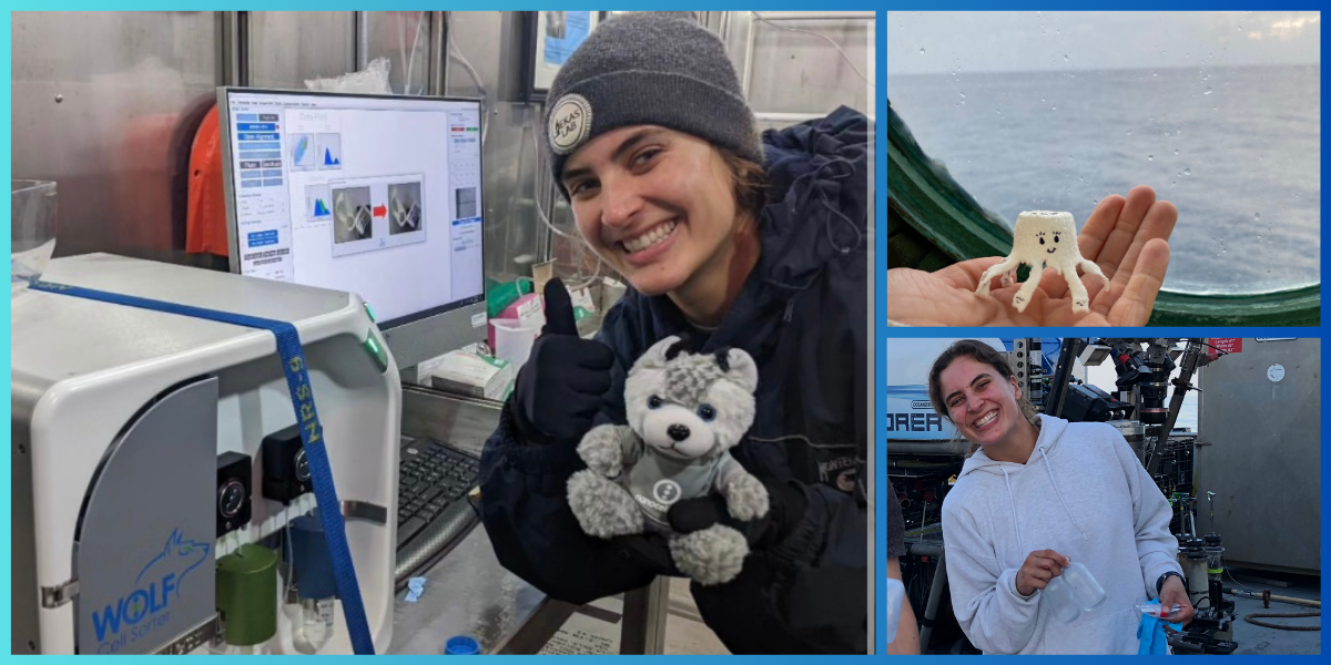
In this case, technology is the easiest solution to healthy cells—NanoCellect’s WOLF G2 Cell Sorter was created to solve this exact problem. Its design was intended to sort cells with:
- Low pressure – An industry-standard flow cytometer machine is set up with far too high of pressures. The WOLF Cell Sorter uses less than 2 psi (which is 30 times gentler than the standard) to ensure that by the time your cells are sorted, you’re not left with an unusable sample.
- High transfection efficiency– With a gentler pressure, the obvious critique was obviously going to speed. However, with the WOLF, you can process 24 microliters of sample per minute. This offers plenty of speed for any lab or research team.
- Disposable cartridges – Plus, for large teams, the disposable cartridges make it easy to switch out samples, while ensuring zero cross-contamination or biohazards being exposed.
To take high-quality pictures, you need a high-quality camera. The same works for cell research. To have a healthy cell population, you need a high-quality cell sorter.
Related:
Next Generation Sequencing and Single Cell Technology
Single Cell Multiomics Analysis
The Homogeneity and Population of Nucleic Acids
There are two parts to this transfection efficiency equation. Optimizing the cell population is one, and optimizing the nucleic acids is the other. For one, you want to ensure that the DNA and RNA you’re introducing into the cell is as close to homogenous as possible. Otherwise, there are too many variables to account for when analyzing the results, and the transfection efficiency will be muddied by different types of genes entering the cell.
Prepare a purified form of nucleic acid chains and then prepare different concentrations to test on cell assays.
For example, by preparing concentrations incrementally from 25-250 ng of nucleic acid to be tested against your cell populations, you can optimize the transfection efficiency per cell type.
- Note: Cell transfection efficiency doesn’t increase with the amount of nucleic acid concentration in a one-to-one fashion. With too much, in fact, you may observe cell toxicity.
Utilizing Correct Cell Population Per Well
When too many cells are bunched up within a single well, you may experience a low transfection rate due to contact inhibition. Contact inhibition is what prevents cells from proliferating without control. It’s why the skin on our bodies doesn’t continue to multiply until we’re all monsters (sorry to make that graphic).
In a well, you don’t want to fill the entire well with the cell population, otherwise you may inhibit some of the regulatory functions of cells: proliferation being one; gene uptake being another.
Attempt to have around 60-80% confluency for optimal transfection efficiency. To set this up, a flow cytometer is used to sort and count cell populations in cell assays.
Improve Your Transfection Efficiency
Whether you work individually in a lab or you’re part of a larger team, having the right transfection technology is a critical component to cell research. And with any of the normal fields-cell biology, cancer research, drug testing, immunophenotyping, etc.-you need a high transfection efficiency. This will not only improve your results in-house, but it will hasten the process from research to publication.
Partnering with NanoCellect means partnering with the best in flow cytometry and cell sorting technology. If you need to improve your transfection efficiency, start by improving your cell populations. The WOLF Cell Sorter is a reliable place to start.
Sources:
- GNN. How are new DNA molecules made? http://www.genomenewsnetwork.org/resources/whats_a_genome/Chp1_4_2.shtml
- Word Counter. How Many Words Are There in the Bible? https://wordcounter.net/blog/2015/12/08/10975_how-many-words-bible.html
- NCBI. Mammalian cell transfection: the present and the future. https://www.ncbi.nlm.nih.gov/pmc/articles/PMC2911531/
- NCBI. Cell toxicity mechanism and biomarker. https://www.ncbi.nlm.nih.gov/pmc/articles/PMC6206310/
- Vanderbilt. Cell Culture Basics. https://www.vanderbilt.edu/viibre/CellCultureBasicsEU.pdf


