Flow cytometry and FACS (fluorescence activated cell sorting) are distinctly different procedures though FACS is a descendant procedure based upon flow cytometry protocols. Advancements in cell sorting technology are contributing in a big way to the molecular science landscape. The overall contributions of what is learned is what guides the pharmaceutical discovery process in both a diagnostic capacity and also therapeutic endeavors which in turn leads to improved patient outcomes1.
Flow cytometry is a method which is used to examine and determine the expression of intracellular molecules and the cell surface and to define and characterize distinct single cell types. It is also used in determining the cell volume, cell size and evaluating the purity of subpopulations which are isolated. This allows the multi-parameter evaluation of individual cells at about the same time.
A flow cytometry is a powerful instrument because it allows simultaneous analysis of both physical and chemical characteristics of up to thousands of particles per second. This makes it a rapid and quantitative method for analysis and purification in cell suspension. Using flow, we can determine the phenotype and function and even sort live cells. i
Some of the measurement applications taken via flow cytometry include:
The two potential flow cytometry data expression formats include:
FACS is an abbreviation for “fluorescence-activated cell sorting”, which is a flow cytometry technique that further adds a degree of functionality. By utilizing highly specific antibodies labeled with fluorescent conjugates (a fluorescent molecule called fluorochrome), FACS analysis allows us to simultaneously collect data on, and sort a biological sample by a nearly limitless number of different parameters. Just like in conventional flow cytometry, forward-scatter, side-scatter, and fluorescent signal data are collected.i
Using a flow cytometer machine, cells or other particles suspended in a liquid stream are passed through a laser beam in single file fashion, and interaction with the laser light is measured by an electronic light detector apparatus as light scatter and fluorescence intensity. If a fluorescent marker is bound to a cellular component, the fluorescence intensity will ideally represent the amount of that particular flow cell component.
Cell sorting via advanced cell sorting instrumentation is performed with high specificity and high standards. Researchers like using the flow cytometry methods because it is the quickest and most effective way to apply measurement to the entire cell process. The operator predetermines or selects certain parameters on how cells should be sorted. i
At this point, the cell sorting machine imposes an electrical charge on each cell so that cells will be sorted by charge (using electromagnets) into separate vessels upon exiting the flow chamber. The technology to physically sort a heterogeneous mixture of cells into different populations is useful for many therapeutic and clinical applications.
To put it the relationship between FACS analysis and flow cytometry in simple context, they are basically partners in the cell analysis process. FACS is used as a cell sorter and enriched for a subset of cells which is often then studied in further detail using flow cytometry or other analytical techniques2.
Flow cytometry is used for cell analysis and is focused on measuring protein expression or co-expression within a mixed population of cells. Both Flow cytometry and FACS are developed to differentiate cells according to their optical filters.
The key distinction between the two cell characterization modalities lies in the functionality of the method being utilized.
Both Flow cytometry and FACS tend to be used interchangeably. They are both developed to differentiate cells according to their optical properties. However, there are some differences in methodology that are distinct and have different procedural outcomes. Used singularly the outcomes are unilateral, and used together, the results are expression data from multiple cell parameters3.
–FACS is a process by which a sample mixture of cells is sorted according to their light scattering and fluorescence characteristics into two or more containers.
–Flow cytometry is a methodology which is utilized during analysis of a heterogeneous population of cells according to different cell surface molecules, size and volume which allows the investigation of individual cells.
-FACS together with flow cytometry can measure and characterize multiple cell generations by using highly specific antibodies tagged with fluorescent dyes, a researcher can perform FACS analysis and simultaneously gather expression data and sort cell samples by a number of variables.
Immunophenotyping: One of the most common application formats used is immunophenotyping. This is a methodology that identifies and quantifies multiple populations of cells in one heterogeneous sample. This includes peripheral blood, bone marrow, and lymph material. Immunophenotyping is primarily used in hematological settings to diagnose certain cancers i.e. malignant lymphoma and/or leukemia4.
Cell Sorting: Advancements in cell sorters are geared towards the gentle isolation of cells into separate tubes. Specialized flow meters with the ability to interrogate and characterize each cell as it passes through the laser and express crucial measurement data of the cell. The key here is also measured in a quantitative sense – the instrumentation is able to handle large volumes that make microscopic examination obsolete.
Cell Cycle Analysis: With flow cytometry and FACS, the cell can be analyzed and measured in all four distinct phases of the entire cell cycle. The cell-based assays can then assist with the determination of cell anomalies using certain fluorescent dyes. The field of single-cell genomics utilizes both of these methods to study a single cell type for a cell population.
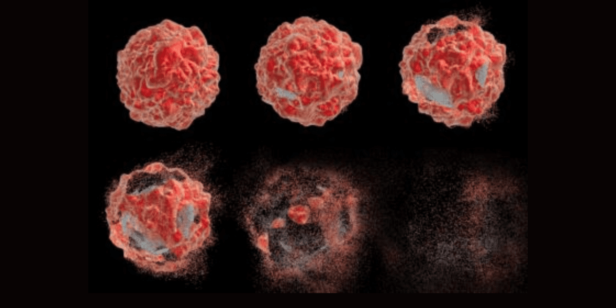
Apoptosis: Apoptosis, or programmed cell death, is a normal part of the life cycle of eukaryotic cells. Cells die for a variety of reasons: through necrosis, brought on by external physical and chemical changes to the cell or through apoptosis, a process in which cells initiate a “suicide” program through internally controlled factors. These two distinct types of cell death, apoptosis and necrosis, can be identified via flow cytometric methodology and used to determine morphological, biochemical and molecular changes occurring in dying cells.
Cell Proliferation Assays: Cell-based assays are one of the key tools to measure mechanism of action responses to certain stimuli such as growth factors, cytokines and other media components. The flow cytometer can measure proliferation by labeling resting cells with a cell membrane fluorescent dye, carboxyfluorescein succinimidyl ester (CFSE). When the cells are activated, they begin to proliferate and undergo mitosis. As the cells divide, half of the original dye is passed on to each daughter cell. By measuring the reduction of the fluorescence signal, researchers can calculate cellular activation and proliferation.
Intracellular Calcium Flux: Cells interact commingle within their environment through signal transduction pathways. When these pathways are activated, membrane-bound calcium ion channels pump calcium into the cell and rapidly increase the intracellular calcium concentration. The higher calcium levels provide the cell with energy to respond. The flow cytometer is able to detect the flux of calcium into the cell and provide detailed measurement data5.
The expansion of flow-cytometric techniques to evaluate intracellular characteristics is moving this platform into the arena of determining cell function. These newer approaches are expanding the utility of flow cytometry as a valuable tool for a deeper understanding of immune function and the role of certain cells as it starts to reveal indicators of apoptosis. By improving call viability through improved technology and recognizing cell death or apoptosis, the technology serves to help researchers understand why it happens and the biochemical changes that occur specific to each phase of cell activity. Charting the signal pathways and DNA fragmentation is a technique that molecular geneticists are finding very valuable. mRNA based therapeutic solutions seem to be key in solving some of the riddles associated with systemic disease6.
As we move closer to being able to exploit the cell analytical process via 3D technology, it is easy to see how the knowledge gained will lead to greater innovation. The research community is looking forward to stretching the blanket of molecular science to include stem cell technology. With stem cells and the ability to clone them effectively and introduce them to the body to create system change is where the future is going. We can see how flow cytometry and the different methodology of FACS will eventually result in more positive patient outcomes, therapeutic interventions, and maybe one- day complete lysis of cancerous tumors. Genetic disease is quickly becoming uncovered and the display of many novel discoveries have started a new movement of biological and molecular science7.
Sources:
2 2https://www.sciencedirect.com/science/article/abs/pii/0022175981902532
3 https://sciencing.com/understand-flow-cytometry-results-5805206.html
4 https://www.cancer.gov/publications/dictionaries/cancer-terms/def/immunophenotyping
5 https://journals.sagepub.com/doi/abs/10.1177/1087057104264038


