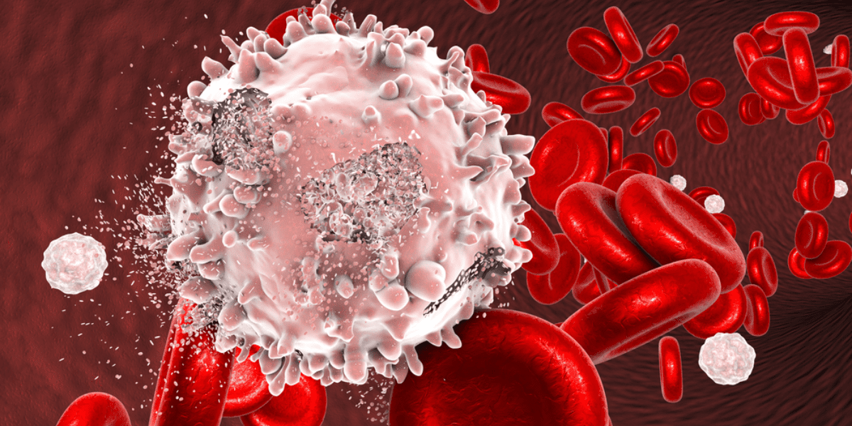
Lymphoma, leukemia, and myeloma are three types of major blood cell cancers. To determine whether a patient is suffering from one of these, a sample of blood is drawn and analyzed under one of two methods:
Both use immunophenotyping to differentiate blood cells and analyze the results to determine if there are cancer cells within the sample. In this article, the former method will be examined as it relates to determining an individual cell’s immunophenotype.
While cancer tests are not its sole purpose, it is one major application that shows why flow cytometry is a powerful tool in cell biology.
Immunophenotyping, in its most basic form, is the technique or method of studying protein expression of cells.
Within cancer testing and research specifically, immunophenotyping consists of using antibodies to label white blood cells and then running the fluorescently labeled cells through a flow cytometer. The FSC and SSC results (forward scatter; side scatter) can detect subtle differences in healthy and cancerous blood cells.
A flow cytometry test via a flow cytometry machine not only differentiates between the types of cancer cells, but also can quantify the cells to determine severity or aggressiveness of the cancer.
An important distinction about the basic principles of flow cytometry is its practicality.
When performing any real-world test (as opposed to theoretical biology), rarely will you work with a homogenous sample. Blood, saliva, bone marrow—these aren’t singular substances. Instead, they’re heterogeneous fluids that contain large quantities of different cells. Flow cytometry grants an automated way to sort, isolate, and quantify these differing cells.
In fact, flow cytometry is used as a tool to streamline the CRISPR process. To learn more about what is CRISPR technology, visit our article explaining the process.
In a paper, Standardizing immunophenotyping for the Human Immunology Project, Maecker writes:
The study of cells moving in suspension through an image plane — flow cytometry — is a potent tool for immunology research and immune response monitoring. Its main advantages are that it makes multiparameter measurements and that it does so on a single-cell basis. The result is that this technique can dissect the phenotypes and functions of cell type subsets in ways that are not possible using a bulk assay, such as Western blots, microarrays or enzyme-linked immunosorbent assays (ELISAs). Nowhere has this proven more useful than in a mixed suspension of immune cells, such as the blood. Newer instrumentation allows for the analysis of eight or more parameters at flow rates of thousands of cells per second; the resulting rich data sets are unparalleled for the knowledge of immune function that they have contributed.
This is high praise for a technique (and machine: the flow cytometer) that has been iteratively improved upon since 1953. Let’s break down what Maecker said to see exactly why it’s such an effective tool for immunophenotyping.
To begin, this test stands out as an exceptional method of understanding cell characteristics because of the multiparameter measurements that are possible. Immune system cells, including an abnormal cell, can be differentiated by looking at the cell surface of proteins. However, with the number of overlaps of these proteins, a method is needed to do multiple measurements simultaneously. To understand the basics behind this, the three main parameters are:
Putting these parameters together allows for an incredible amount of application across different biological and medical fields.
Imagine trying to study a cluster of microscopic cells by running them through a laser to measure their light refraction. Unless you’re isolating each cell individually, the practice is moot. There’s variability within individual cell populations, for example in blood…
Without proper detection and analysis methods, these values quickly derange any hope of flow cytometry lab tests being practical (that word, again, is important). Secondly, if there was no control over the measurement system, these monocytes and lymphocytes would be streaming through the laser simultaneously, not to mention the other neutrophils, eosinophils, red blood cells, and the cancerous cells joining in on the party. The light scattering in all directions would create a cohesive slab of chaos.
There’s only one solution, the cells have to pass the laser one at a time.
See the problem here?
Cells are on a microscopic level. There’s no manual way to line them up single file. Instead, properties of fluid dynamics are utilized to achieve this.
Depending on the pressure of the core stream, this will help to negate doublets (when two cells run through the laser simultaneously). The lower pressure systems are slower, though they tend to work more efficiently.
The cells are passing through the laser one at a time. Their light refraction is being measured using forward scatter and side scatter, and the fluorescent labeled dyes help isolate certain cell populations. Now it’s time to determine the phenotypes and functions of the cell subsets. These properties can be ascertained using:
Performing a flow cytometry blood sample test, or frankly any flow cytometry lab test, will involve a heterogeneous solution. In the case of blood, you are going to have a mixed suspension of various immune cells that would otherwise not be able to stay separated.
Apart from being able to measure multiple parameters at once, Maecker claims that the newer instrumentation can analyze eight or more parameters. Question is, what kind of parameters are these (besides the ones mentioned above), and how are they used?
The word, again, to come back to is practical. Flow cytometers can perform all of the above functions and provide invaluable data across multiple fields at an incredible speed. Thousands of cells per second can flow through the laser, be recorded for analysis, and then be sorted for further testing.
This is where flow cytometry strides ahead of any other technique that is available for these types of measurements.
Of course, with the ever-advancing landscape of technology, certain flow cytometers will be more efficient at immunophenotyping than others. If you want a machine that will perform flow cytometry lab tests with incredible efficacy, there’s NanoCellect’s WOLF Cell Sorter.
Not only will you be able to determine a cell’s immunophenotype, but you can do so without worrying about killing a chunk of your sample cells in the process. The WOLF Cell Sorter was designed to be ten times more gentle on cells than the next leading gentle flow cytometer. This provides the opportunity to continue testing after your cells have been sorted.
At NanoCellect, we know happy cells.


