How Flow Cytometry is Used to Study Infectious Disease

Flow cytometry has been around for decades and has contributed to vital research on deadly diseases like cancer, malaria, tuberculosis, and HIV, both in the lab and out in the field. Not only does it help advance known treatments and therapies for various chronic diseases, but it also helps understand and develop treatment for emerging infectious diseases.
So how does flow cytometry relate to a hot topic like infectious disease?
And how exactly is flow cytometry used to study infectious disease?
Read on for more on how the cell sorter works, how it’s used for infectious disease research, and which devices are best for the process.
What is a Flow Cytometer?
It’s a device that performs the following actions to cells suspended in a solution:
- Analyze individual cells from a mixture.
- Quantitate cells using laser optics and fluorescent markers.
- Sort individual cells flagged by key properties (if a cell sorting feature is included).
When analyzing cells, flow cytometry analysis can identify quite a few features, like:
- Size
- Granularity
- Specific surface receptors
- Intracellular proteins
- Gene expression
- Total DNA
- Newly synthesized DNA
- Transient signaling events
During its long tenure as a research instrument, flow cytometers have typically been big and cumbersome—until recently, that is. Some newer models have become far more efficient, portable, and easy-to-use thanks to innovation and advancements in the field. But more on that later.
How Does a Flow Cytometer Work?
You may be wondering how exactly this device accomplishes such outstanding analytical feats on such a tiny scale.
The secret? A mix of fluidics, laser optics, light detectors, fluorescence, and software.
Here’s what happens when you place a cell suspension in a flow cytometer:
- The agitated cell suspension is shot into a saline solution stream.
- Due to hydrodynamics and the narrowing geometry of the fluid channel, the cell suspension is compelled to form a single-file line of individual cells.
- This line then passes through a laser beam, reflecting and refracting light off each cell into forward scatter (FSC) and side scatter (SSC) or back scatter (BSC).
- Precise optical devices guide the FSC and SSC/BSC into their respective detectors, which convert light readings into a voltage pulse, which are then read by the computer processing unit (CPU), interpreted for analysis, and displayed on the screen.
But how does scatter help you to analyze cells? These light measurements indicate different properties of the cell, such as:
- Forward Scatter – Read from a detector in the immediate laser path, FSC readings are directly proportional to the size of a cell.
- Side Scatter/Back Scatter – Read from the detector perpendicular to or behind the laser path, SSC/BSC determines the shape of a cell, as well as some of its internal features.
By combining these two readings, researchers can now distinguish between different cell types in a mixture. This identification allows them to label, separate, and sort them for further review.
Using Fluorescence for Analysis
To identify further cell properties in a sample, researchers can also stain or dye cells with fluorophores: fluorescent markers that can be excited by and emit specific wavelengths of light.
These markers bind to different components that researchers may want to identify, such as:
- DNA
- RNA
- Certain proteins
- Red blood cells
- White blood cells
A flow cytometer can then detect the light emitted by the fluorophores in each cell and based on their intensity, measure the relative amount of that specific component. This procedure is called immunophenotyping, which is crucial for disease diagnosis and prognosis.
The more fluorophore colors your flow cytometer allows you to use, the more components you can identify at once.
Applications of Flow Cytometry
This fascinating technology has been used for years in a variety of applications. In one notable application, it has identified how many CD4+ versus CD8+ T lymphocytes there are in blood samples to discern whether an HIV infection has led to AIDS, and to what degree anti-HIV medication is working.
In addition to this noble cause, flow cytometry can also do the following:
- Perform blood counts
- Observe different leukocyte types
- Determine total DNA per cell in tumor biopsies when diagnosing and treating cancer
- Sort T cells to determine how an infection has affected their function
Flow cytometry is essential in infectious disease research and treatment. It allows for a simple and fast way to study, diagnose, identify spread, and inform appropriate treatment strategies.
How Researchers Use Flow Cytometry to Study Infectious Disease
With infectious diseases present in overwhelming numbers—AIDS and tuberculosis alone are responsible for 4.5 million deaths annually—affordable, efficient, and reliable immunophenotyping and cell isolation are vital in the battle for prevention, treatment, and eradication.
With new microbial pathogens constantly springing up from all corners of the world, flow cytometry, more than ever, is fundamental in controlling the spread of a viral infection.
Here are the main ways flow cytometers assist in the treatment and study of infectious disease:
- Immunophenotyping allows researchers to diagnose quickly and accurately.
- Effectiveness of emerging treatments and therapies can be tested rapidly and iterated until a solution is found.
- Cells can be sorted to study the specific effect that the infection has on them.
- Pathogenic microbes can be detected from environmental samples to control spread, track and predict proliferation.
Flow cytometry enables public health research with these advantages:
- Precision – According to a publication by Dr. Babetta Marrone (Ph.D.) from the Los Alamos National Laboratory, “measurements made at the level of individual cells… provide a discrete observation that is not achievable by bulk analysis methods, which average results across all cells in the sample… The high sensitivity is achieved by the inherent ability of flow cytometry to resolve free from bound fluorescence by virtue of the small analysis volume used.”
- Speed – Thanks to high throughput and “multi-tasking” abilities, flow cytometers can analyze enormous amounts of unique components per sample tube.
- Versatility – With the continued development of fluorophores and reagents, flow cytometers can measure even more components like lipids, proteins, RNA, and DNA. They can perform a range of tasks, like characterizing host-cell susceptibility and immune response or identifying viruses and bacteria.
- Robust Analysis – Due to their ability to process tens of thousands of cells per second, flow cytometers allow researchers to produce statistics with huge sample sizes, resulting in vast amounts of rapid data to be used towards the eradication of infectious disease.
Biosecurity – measures taken to stop the introduction or spread of harmful organisms – applications for flow cytometry are as follows:
- Measuring and predicting the threat of harmful pathogens through lab and fieldwork while addressing biosafety concerns.
- Rapidly detecting biological threats through environmental surveillance.
- Analysis of how pathogens damage and proliferate within human hosts.
- Monitoring immune response in infected hosts.
By quickly identifying biological threats and pathogens in an efficient way, flow cytometers allow researchers to effectively tackle multiple infectious diseases, by testing for a versatile array of characteristics—saving lives along the way.
Sort Cells Gently With the WOLF
Flow cytometers perform this incredibly detailed analysis in a matter of minutes. Impressive. However, the quality of such testing is based on several factors, including:
- Ease-of-use
- Portability
- Sample-to-sample contamination
- Signal noise
- Cell death
A bulky, difficult-to-use flow cytometer can result in misuse by lab personnel and poor results. A poorly designed cell sorting feature can lead to contamination between samples, thereby wasting valuable cells and ruining important results.
Most importantly, a flow cytometer that exerts too much pressure can damage cells and reduce the viability of a cell population. Dead cells can become distracting noise in data analysis or result in complete experimental failure if the cells are sorted. When it comes to the crucial and precise act of single cell analysis and sorting, cell death is the last thing you want.
That’s where the WOLF Cell Sorter comes in.
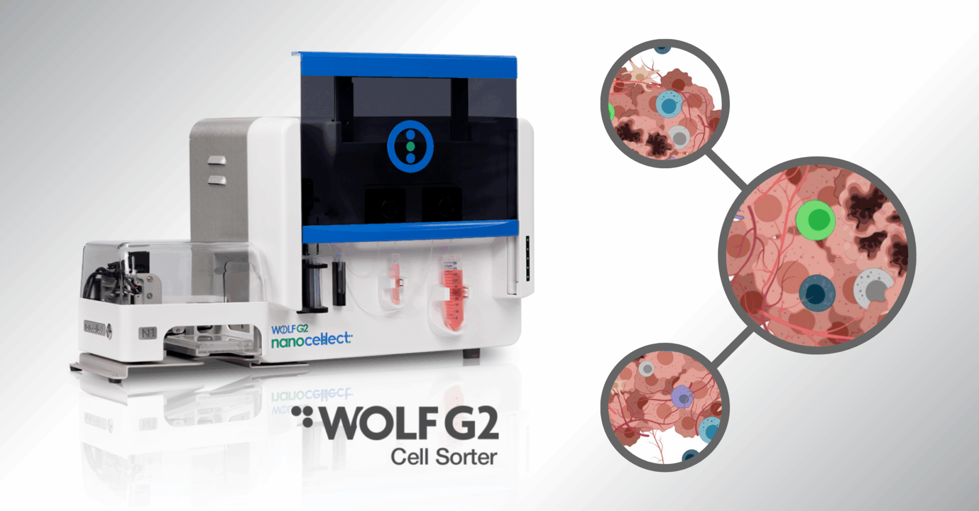
NanoCellect’s WOLF-G2® Cell Sorter and N1 Single-Cell Dispenser mitigate these common threats against successful experiments through their superior design and intuitive software. Thanks to innovative engineering, researchers no longer have to worry about cell death, contaminated samples, or complicated instrumentation.
Here’s how it stands out:
- The WOLF uses a sorting pressure of <2 psi, which is about thirty times lower than the competition, resulting in far fewer dead cells.
- High-performance PMT detectors can spectrally filter fluorophores into three different colors for simultaneous analysis.
- Its piezoelectric sorting cartridges are gentle on cells, sterile, and disposable, to prevent cross-contamination and preserve cell samples.
- The WOLF’s intuitive, easy-to-use software is accessible to researchers of every experience level, allowing beginners to adapt quickly, and experts to perform a multitude of complex analyses.
- The patented cell-sorting technology allow researchers to sort two populations at the same time.
For single cell multiomics and sorting, choose a device that’s gentle, simple, and reliable. With the WOLF G2 Cell Sorter you get happy cells and better science.
Sources:
- Janossy, George. Howard Shapiro. Simplified Cytometry for Routine Monitoring of Infectious Diseases. https://pubmed.ncbi.nlm.nih.gov/18228555/
- Marrone, Babetta. Flow Cytometry: A Multipurpose Technology for a Wide Spectrum of Global Biosecurity Applications. https://journals.sagepub.com/doi/full/10.1016/j.jala.2009.03.001
- MIT StarCellBio. Flow Cytometry Animation (Video). https://www.technologynetworks.com/cell-science/videos/how-does-flow-cytometry-work-317076
- Mass. Amherst Microbiology 542. What is Flow Cytometry
- https://www.bio.umass.edu/micro/immunology/facs542/facswhat.htm


