Image-Based Microfluidic Cell Sorting Using Epi-Fluorescence Microscope Configuration

Abstract
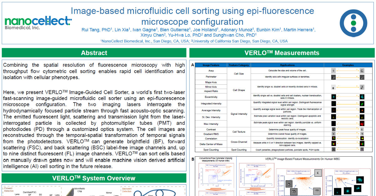
Combining the spatial resolution of fluorescence microscopy with high throughput flow cytometric cell sorting enables rapid cell identification and isolation with cellular phenotypes. Here, we present VERLOTM Image-Guided Cell Sorter, a world’s first two-laser fast-scanning image-guided microfluidic cell sorter using an epi-fluorescence microscope configuration. The two imaging lasers interrogate the hydrodynamically focused particle stream through fast acousto-optic scanning. The emitted fluorescent light, scattering and transmission light from the laser-interrogated particle is collected by photomultiplier tubes (PMT) and photodiodes (PD) through a customized optics system. The cell images are reconstructed through the temporal-spatial transformation of temporal signals from the photodetectors. VERLO can generate brightfield (BF), forward scattering (FSC), and back scattering (BSC) label-free image channels and, up to nine distinct fluorescent (FL) image channels. VERLO can sort cells based on manually drawn gates now and will enable machine vision derived artificial intelligence (AI) cell sorting in the future release.
PST-014
